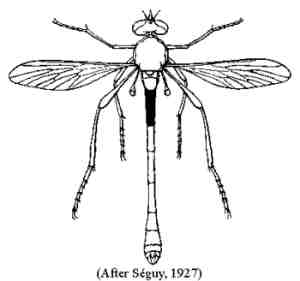
Abdominal tergite 1 five or more times as long as wide (Fig. 1). Alula and pulvilli lacking. Abdominal sternite 1 extending about halfway back under tergite 2
|
Abdominal tergite 1 no more than four times as long as wide. Usually both alula and pulvilli present. Abdominal sternite 1 confined beneth tergit 1
|
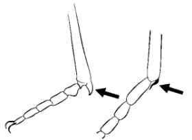
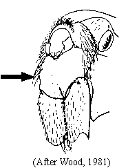
Fore tibia with an apical spur; one of the spines at the apex of the ventral side of the fore tibia differentiated, enlarged and stouter than remaining spines, or if not noticeably larger, twisted and sigmoid (Fig. 2). Prosternum dissociated by a membranous area from proepisternum, except in Blepharepium Rondani (Fig. 3)
|
Fore tibia without an apical spur; all apical spines on fore tibia straight, or if one is slightly curved then it is not thickened or sigmoid. Prosternum either dissociated from proepisternum or fused to it
|
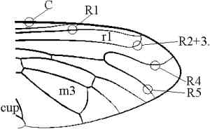
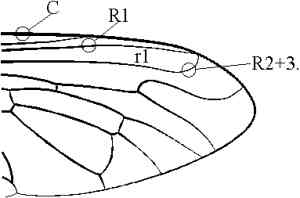
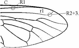
Apex of R2+3 directed sharply forward, meeting C at an angle of about 90°, ending either at distal end of R1 (cell r1 closed) (Fig. 4) or a short distance from R1 along C (cell r1 open) (Fig. 5). Vein R4 strongly sinuate and arched forward after separation from R5. Cells m3 and cup always closed before wing margin. Prosternum fused to proepisternum (Fig. 6). Male with only six tergites visible dorsally
|
Apex of R2+3 not directed sharply forward before ending in C or R1. R4 not unusually arched and sinuate. Cells m3 and cup open to wing margin, or one oth the two closed, or both closed. Prosternum dissociated from proepisternum or fused to it. Male with six to eight tergites visible dorsally
|
R2+3 ending in C (except in Enigmomorphus Hermann) (Fig. 7). Neither a strong bristle present on the supero-posterior angle of anepisternum nor a row of bristles present on the katatergite (Fig. 8)
|
R2+3 joining R1 proximal to end of R1, with cell r1 thus separated from wing margin (Fig. 9). Either anepisternum with at least one strong bristle on its supero-posterior angle, or katatergite with a vertical row of bristles or bristly hairs (Fig. 8)
|
||||
|
|||||
Prosternum dissociated from proepisternum by a membranous area (Fig. 3)
|
Prosternum fused to proepisternum, forming a precoxal bridge (Fig. 6)
|
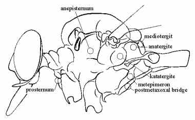
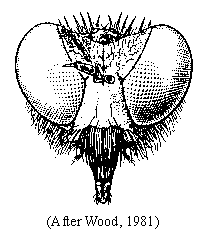
Frons narrowed at level of insertion of antennae and then suddenly and widely diverging towards apex, which is ectremely shallow (eyes much more distant at vertex than at antennal level) (Fig. 10). Face without tentorial pits or grooves, flat above and prominent below or very gibbose. Posterodorsal corner of metepimeron bare (Fig. 8). Abdomen slender. Female terminalia with characteristic ventral keel and spines (Fig. 11)
|
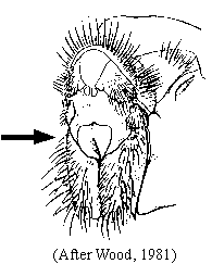
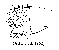
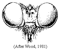
Frons approximately of same width at level of antennal insertion and vertex, the latter excavated (eyes not noticeably more distant at vertex then at antennal level) (Fig. 12). Face with pronounced tentorial pits or grooves extending well above lower facial margin. Face in profile not produced beyond eye margin. Posterolateral corners of metepimeron with short hairs (Fig. 8). Abdomen very short, usually three-quarters or less width of wing. Female terminalia simple, tubular, without spines (Fig. 13). Remark: Some genera seems to have spines on acanthophorites and a keel on sternite 8 in females (e.g. Oligopogon)
| ||||||||
|
|||||||||
Supero-posterior angle of anepisternum, infront of wing insertation, with at least one strong, long bristle. Katatergite never with vertical row of bristles (Fig. 8). Prosternum fused to proepisternum (Fig. 6). Palpus, one or two segmented. Female terminalia without spines
|
Supero-posterior angle of anepisternum never with a strong, long bristle. Katatergite with a vertical row of long bristles or bristly hairs (rarely reduced to only one bristle) (Fig. 8). Prosternum dissociated from proepisternum or fused to it. Palpus always one-segmented. Female terminalia with or without spines
|
Anatergite pilose, the hair situated on top of it (Fig. 8). R4 never with an extra vein
|
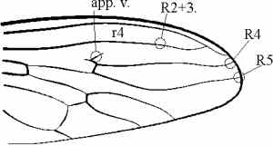
Anatergite bare, or if some hairs, these placed mostly on latero-internal margin of anatergite and on immediately adjacent area of mediotergite, but never on top of anatergite and R4, in this case , always with a short extra vein present at its junction with R5, the short vein ending in cell r2+3 (Fig. 9)
|
Antennal style plumose (Fig. 15). Postmetacoxal area heavily sclerotized, forming a complete bridge behind hind coxae (Fig. 16)
|
Antennal stylus bare. Postmetacoxal area membranous*
|
![]()
![]()
Last saved June 07 2003
© F. Geller-Grimm