Note 1: After comparison between individuals from french continent [Le Grau-du-Roi, department of Gard, on the beach, 22.06.1997 and published by G. Tomasovic, 1998, Notes sur les Asilidae Paléarctiques (Diptera, Brachycera) 4-8, Bull. Annls Soc. r. belge Ent. 134 : 133-138. As Andrenosoma leucogenys Séguy, 1952] and individuals from Corsica, personnal catches (Oriental beach, Ghisonaccia, 23.07.1997 and Aleria, 12.07.2000) and one male specimen from the British Museum, sended by Dr.J.Chainey, from also Corsica(Occidental beach, F. Guglielmi, Ajaccio, 20.08.1906) and identified by H. Oldroyd as Andrenosoma albopilosum Villeneuve, 1911. All this specimen belong to Andrenosoma albopilosum Vill., their dististylus are identicals and the Seguy's drawing Nº2(p. 195) in his paper [1952, Andrenosoma nouveaux de France, Rev. Fr. d'Ent., 19 : 192-196] isn't the dististylus of A. leucogenys but that's of albopilosum; it's certainly a mistake. Unfortunately the "type", one unique specimen of A. leucogenys, I can't find it in the entomologicals collections in Paris! Very disturbing!!
Jean-Michel MALDES, maldes@cirad.fr, CIRAD-Amis - Protection des Cultures, TA 40 / L - Campus International de Baillarguet - CSIRO, 34398 Montpellier Cedex 5 (FRANCE)
Note 2: Mike Taylor is working on Andrenosoma cornuta Oldroyd, 1972. If you have material from Greece and Turkey, please get in touch with him. It seems to be that a new desription of this species is necessary. His address is:
18 Albany Road, Bramhall, Stockport, Cheshire, SK7 1ND, United Kingdom, mike.taylor@tiscali.co.uk
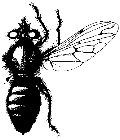
A. atra (copied from Goot, 1985)
| [Without consideration: A. serratum Hermann, 1906; A. trigoniferum Hermann, 1906; A. violacea (Fabricius, 1781)]. | ||
| 1 | 1st posterior cell closed and stalked on margin (Fig. 2); tergites black or dark violet (male genitalia Fig. 5ab). Distribution: Europa: A / BG / CH / CS / D / DK / E / F / GR / I / PL / S / YU / USSR / CET / SET / North Africa / Algeria |
atra (Linnaeus, 1758) |
| - | 1st posterior cell open on margin (Fig. 3) or closed before wing-margin (Fig. 4). | 2 |
| 2 | Ocellar tubercle conspicuously raised as a three-pronged structure, 3rd antennal segment with longitudinal fissure on distal quarter (figures). Distribution: Asia: Turkey & Europe: GR [unpublished, record: M. Taylor] |
cornuta Oldroyd, 1972 |
| - | Ocellar tubercle without three-pronged structure. | 3 |
| 3 | 1st posterior cell closed (or nearly closed) on margin (Fig. 4). | 4 |
| - | 1st posterior cell open on margin (Fig. 3). | 5 |
| 4 | Tergites red in the middle (male genitalia Fig. 7). Distribution: Europa: F |
bayardi Séguy, 1952 |
| - | Tergites black, hind lateral corners of most segments white pruinose; mesonotum with white stripes. Distribution: Europa: GR |
pusillum Hermann, 1906 |
| 5 | Tergites black. | 6 |
| - | Tergites red in the middle; body with white and black setae. | 7 |
| 6 | Mystax white; body with yellow-white setae and brownish bristles; predominant white setae on legs; wing venation brown (male genitalia Fig. 9). Distribution: Europa: F (Corsica) |
albopilosum Villeneuve, 1911 |
| - | Mystax black above lower facial margin; short setae on mesonotum black; legs with white and black setae; wing venation black (male genitalia Fig. 10). Distribution: KMA |
jenisi Kovár & Hradský, 1996 |
| 7 | Setae of face white only; setae of mesonotum long and white (male genitalia Fig. 11). Distribution: Europa: F |
leucogenys Séguy, 1952 |
| - | Setae of face white and black; mesonotum with short setae. | 8 |
| 8 | Tergites 2 - 7 orange in the middle (male genitalia Fig. 12). Distribution: Europa: A / CS / D / E / F / GR / I / PL / R / TR / USSR / CET / SET / ES / FE / Asia / Turkey / [ / CH / ] |
albibarbe (Meigen, 1820) |
| - | Tergites 4 - 7 orange in the middle only (male genitalia Fig. 13). Distribution: Europa: I / F |
cyrtoxys Séguy, 1952 |
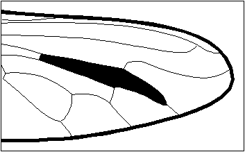
Fig. 2: Wing of A. atra, 1st posterior cell closed and stalked.
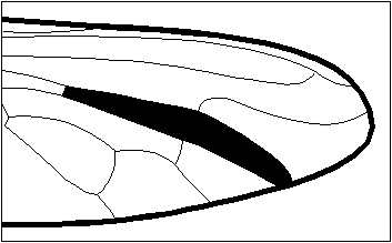
Fig. 3: Wing of A. albibarbe, 1st posterior cell open.
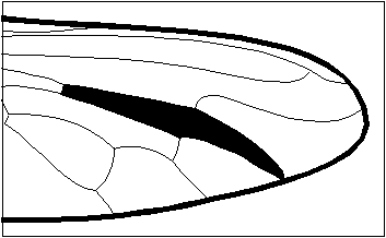
Fig. 4: Wing of A. bayardi, 1st posterior cell closed before margin.

Fig. 5ab: A. atra male genitalia (a. copied from Séguy, 1952; b. copied from Kovár & Hradský, 1996).
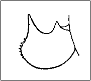
Fig. 7: A. bayardi male genitalia (copied from Séguy, 1952).
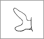
Fig. 9: A. albopilosum male genitalia (copied from Séguy, 1952).

Fig. 10: A. jenisi male genitalia (copied from Kovár & Hradský, 1996).

Fig. 11: A. leucogenys male genitalia (copied from Séguy, 1952).
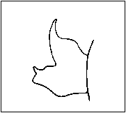
Fig. 12: A. albibarbe male genitalia (copied from Séguy, 1952).
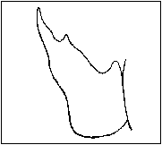
Fig. 13: A. cyrtoxys male genitalia (copied from Séguy, 1952).
Last saved March 11 1999
© F. Geller-Grimm