 Introduction
 History
 Morphology
 Terminology
 Head
Head
 Antenna
 Thorax
 Leg
Leg
 Wing
Wing
 Abdomen
 Genitalia
 Chaetotaxy
 Egg
Egg
 Larva
Larva
 Pupa
Pupa
 Addition
 ESEM
ESEM
 Glossary
 Phylogeny
 Distribution
 Ecology
 Biology
 Collecting
 Determination
 Photography
 References
|
|

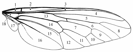
Fig. 1: wing, cells - Ceraturgus
| |
Fig. 1:
1 = basal costal cell, bc [1/2]. -
2 = costal cell, c [1/2]. -
3 = subcostal cell, sc [1/2]; mediastinal cell [2]. -
4 = 1st radial cell, r1 [1]; marginal cell [2/3]. -
5 = 2nd + 3rd radial cell, r2+3 [1]; 1st submarginal cell [2/3]. -
6 = 5th radial cell, r5 [1]; 1st posterior cell [2/3]. -
7 =discal cell, 1m2 [1/2/3]. -
8 = 4th radial cell, r4 [1]; 2nd submarginal cell [2/3]. -
9 = 1st medial cell, m1 [1]; 2nd posterior cell [2/3]. -
10 = 2nd medial cell, m2 [1]; 3rd posterior cell [2/3]. -
11 = 3rd medial cell, m3 [1]; 4th posterior cell [2/3]. -
12 = anterior cubital cell, cua1 [1]; 5th posterior cell [2]. -
13 = basal radial cell, br [1]; 1st basal cell [2]. -
14 = basal medial cell, bm [1]; 2nd basal cell [2]. -
15 = posterior cubital cell, cup [1]; 3rd basal cell, anal cell, lower basal cell [2]. -
16 = anal cell, a1+2 [1]; axillary cell [2]. -
17 = alula [1/2/3]; axillary lobe, posterior lobe [2]. -
18 = intervenal area [5].
|

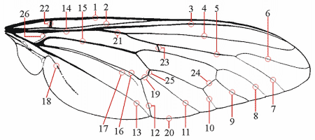
Fig. 2: wing, veins - Ceraturgus
| |
Fig.2:
1 = costa, C [1/2]. -
2 = subcosta, Sc [1/2]; auxiliary vein [2]. -
3 = anterior branch of radius, R1 [1]; 1st branch of radius, 1st longitudinal vein [2]. -
4 = 2nd and 3rd posterior branch of radius, R2+3 [1]; 2nd longitudinal vein [2]. -
5 = 4th and 5th posterior branch of radius, R4+5 [1]; 3rd longitudinal vein [2]. -
6 = 4th posterior branch of radius, R4 [1]; anterior branch of 3rd vein [2]. -
7 = 5th posterior branch of radius, R5 [1]; posterior branch of 3rd vein [2]. -
8 = 1st posterior branch of media, M1 [1]; 1st branch of medius, 4th vein [2]. -
9 = 2nd posterior branch of media, M2 [1]; 2nd branch of 4th vein, anterior intercalary vein [2]. -
10 = 3rd posterior branch of media, M3 [1]; 3rd branch of 4th vein, posterior intercalary vein [2]. -
11 = 1st anterior branch of cubitus, CuA1 [1]; 1st branch of cubitus, 5th vein [2]. -
12 = 2nd anterior branch of cubitus, CuA2 [1]; 2nd branch of 5th vein [2]. -
13 = 1st branch of anal vein, A1 [1]; 2nd anal vein [2]. -
14 = radius, R [1]; main stem of radius [2]. -
15 = media, M [1]; main stem of medius [2]. -
16 = cubitus, Cu [1]; main stem of the cubitus [2]. -
17 = posterior branch of cubitus, CuP [1]; 1st anal vein [2]. -
18 = 2nd branch of anal vein, A2 [1]; remnant, 3rd anal vein [2]. -
19 = anterior branch of cubitus, CuA [1/2]. -
20 = posterior margin of wing, - [1]; ambient vein [2/3]. -
21 = radial sector, Rs [1/2]. -
22 = humeral crossvein, h [1/2]. -
23 = radial-medial crossvein, r-m [1]; anterior small crossvein, anterior middle crossvein [2]. -
24 = medial crossvein, m-m [1/2]; posterior crossvein [2]; [above m-m: base of M2]. -
25 = medial-cubital crossvein, m-cu [1]; discal crossvein; mediocubital crossvein [2]. -
26 = anterior branch of media, MA, brace vein, phragma [1]; arculus [2]. -
|

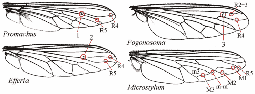
Fig. 3: wing, special venation
| |
Fig. 3:
1 = R4 with recurrent vein arising its junction with R5; recurrent branch extending toward base of wing until it joins R2+3 [4]. -
2 = R4 with recurrent vein arising its junction with R5; recurrent branch short, ending in cell r2+3 [4]. -
3 = R2+3 and R4 connected by a short extra crossvein [4]. -
|
References
[1] McAlpine, J.F. (1981): Morphology and terminology - Adults. - In: McAlpine, J.P. et al. (eds.): Manuel of Nearctic Diptera, vol. 1; p. 9-63 - Ottawa: Research Branch, Agriculture Canada, Monograph 27.
[2] Hull, F.M. (1962): Robber flies of the world, 2 volumes; 907 pp. - Washington: Bulletin of the United States National Museum 224 (1,2).
[3] Theodor, O. (1980): Fauna Palaestina - Insecta II - Diptera: Asilidae; 446 pp. - Jerusalem: The Israel Academy of Science and Humantities.
[4] Wood, G.C. (1981): Asilidae. - In: McAlpine, J.P. et al. (eds.): Manuel of Nearctic Diptera, vol. 1; p. 549-573 - Ottawa: Research Branch, Agriculture Canada, Monograph 27.
[5]Crampton, G.C. (1942): Guide to the insects of Connecticut. Part IV. The Diptera, or true flies of Connecticut. The external morphology of the Diptera. - Bulletin of the Connecticut Geological and Natural History Survey 64: 10-165; Hartford.
|


|